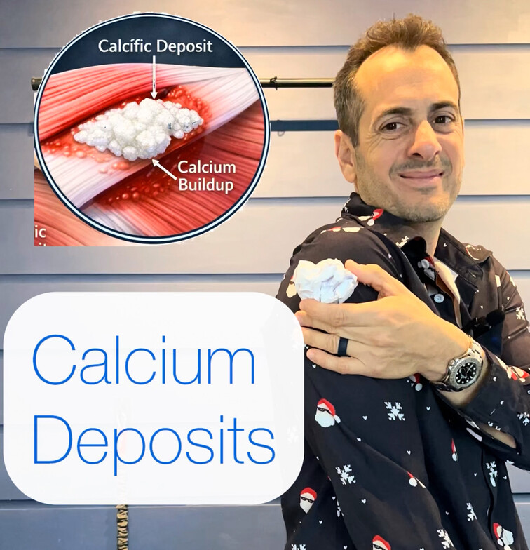
Calcium deposits in muscles and tendons—commonly referred to as calcific deposition—are the result of disrupted tissue healing. They are most often associated with repetitive micro-trauma, chronic mechanical overload, and persistent inflammation rather than acute injury.
When soft tissue is exposed to repeated stress, local blood flow and oxygen delivery can become compromised. This creates a hypoxic environment that interferes with normal cellular repair processes. In healthy healing, fibroblasts and tendon cells lay down organized collagen to restore tissue integrity. However, under chronic inflammatory conditions, this repair response becomes dysregulated.
As a result, calcium hydroxyapatite crystals may accumulate within the soft tissue instead of normal collagen fibers. These deposits increase tissue density and stiffness, disrupt normal muscle and tendon glide, and place increased stress on surrounding structures. Over time, this altered tissue quality can activate local pain receptors, contributing to pain, weakness, and reduced range of motion.
Calcific deposits are most commonly seen in tendons with high mechanical demand and limited blood supply, such as the rotator cuff of the shoulder, but they can occur in other regions exposed to repetitive loading.
Effective management focuses on improving tissue health rather than simply addressing pain. Reducing inflammation, restoring blood flow, improving mobility, and optimizing load management are essential for supporting proper tissue repair and preventing recurrence.

Dr. Steve Muscari
Contact Me